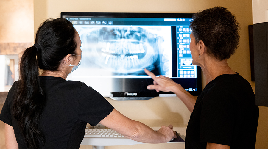
Digital Radiography
Our Doctors choose carefully which and when radiographs are taken. There are many guidelines that we follow. Radiographs allow us to see everything we cannot see with our own eyes. Radiographs enable us to detect cavities in between your teeth, determine bone level, and health of bone. We can also examine the roots and nerves of teeth, diagnose lesions such as cysts or tumors, as well as assess damage when trauma occurs.
Dental radiographs are invaluable aids in diagnosing, treating, and maintaining dental health. Exposure time for dental radiographs is extremely minimal. Our Doctors utilize Digital Imaging Technologies within the office. With digital imaging, exposure time is about 50 percent less when compared to traditional radiographs. Digital imaging can also help us retrieve valuable diagnostic information. We may be able to see cavities better.
The advantages of digital imaging enables us to not only store patient images, but also enables us to quickly transfer them to appropriate specialists or insurance companies.
Digital X-Rays:
Digital X-rays offer more precision since we view the image on a computer monitor, instead of holding up a 35mm film up to the light. Digital X-rays result in 1/6th the radiation exposure to you.
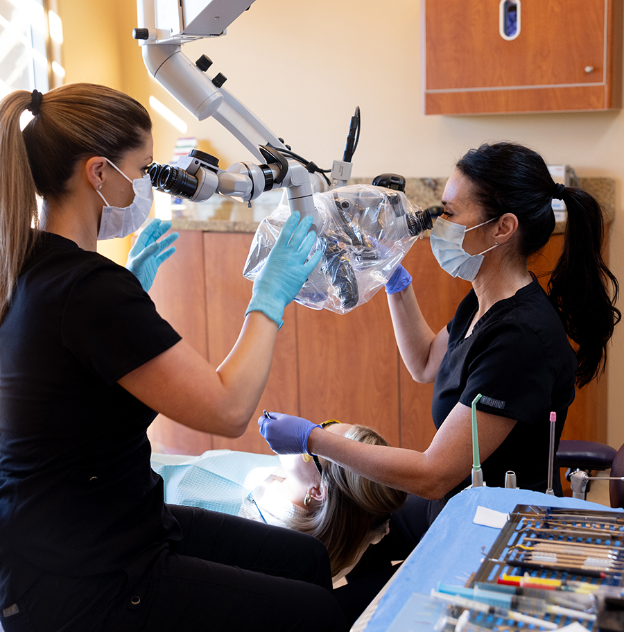
Surgical Microscopes
Magnification and fiber–optic illumination are invaluable tools that assist our doctors in performing the technical aspects of endodontic treatment. Below are just a few applications that our doctors utilize the Surgical Microscope.
- Improved lighting and magnification aid in locating additional canals.
- Improves ability to thoroughly clean prepared canals.
- Aids in retreatment of broken instruments.
- Accurate depth of retro-prep extension can be more easily assessed.
- Improves detection and evaluation of root fractures and abnormalities.
- Allows our doctors to minimize the size of the surgical site, reducing patient discomfort and healing time.
- Improves accuracy of microsurgical incisions and suturing with 6–0 through 8–0 sutures, permitting precise tissue/tissue and tissue/tooth approximation for primary would healing.
- Provides high-resolution video for patient education, enhanced training, and insurance/legal documentation.